Table of contents
Volume 5, Issue 7, pp. 300 - 356, July 2018
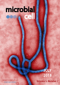
Cover: Digitally-colorized transmission electron micrograph of an Ebola virus virion. Image by Cynthia Goldsmith (Centers for Disease Control and Prevention, USA; Public Health Image Library, image ID #10816); image modified by MIC. The cover is published under the Creative Commons Attribution (CC BY) license.
Enlarge issue cover
Methodologies for in vitro and in vivo evaluation of efficacy of antifungal and antibiofilm agents and surface coatings against fungal biofilms
Patrick Van Dijck, Jelmer Sjollema, Bruno P.A. Cammue, Katrien Lagrou, Judith Berman, Christophe d’Enfert, David R. Andes, Maiken C. Arendrup, Axel A. Brakhage, Richard Calderone, Emilia Cantón, Tom Coenye, Paul Cos, Leah E. Cowen, Mira Edgerton, Ana Espinel-Ingroff, Scott G. Filler, Mahmoud Ghannoum, Neil A.R. Gow, Hubertus Haas, Mary Ann Jabra-Rizk, Elizabeth M. Johnson, Shawn R. Lockhart, Jose L. Lopez-Ribot, Johan Maertens, Carol A. Munro, Jeniel E. Nett, Clarissa J. Nobile, Michael A. Pfaller, Gordon Ramage, Dominique Sanglard, Maurizio Sanguinetti, Isabel Spriet, Paul E. Verweij, Adilia Warris, Joost Wauters, Michael R. Yeaman, Sebastian A.J. Zaat, Karin Thevissen
Reviews |
page 300-326 | 10.15698/mic2018.07.638 | Full text | PDF |
Abstract
Unlike superficial fungal infections of the skin and nails, which are the most common fungal diseases in humans, invasive fungal infections carry high morbidity and mortality, particularly those associated with biofilm formation on indwelling medical devices. Therapeutic management of these complex diseases is often complicated by the rise in resistance to the commonly used antifungal agents. Therefore, the availability of accurate susceptibility testing methods for determining antifungal resistance, as well as discovery of novel antifungal and antibiofilm agents, are key priorities in medical mycology research. To direct advancements in this field, here we present an overview of the methods currently available for determining (i) the susceptibility or resistance of fungal isolates or biofilms to antifungal or antibiofilm compounds and compound combinations; (ii) the in vivo efficacy of antifungal and antibiofilm compounds and compound combinations; and (iii) the in vitro and in vivo performance of anti-infective coatings and materials to prevent fungal biofilm-based infections.
Shepherding DNA ends: Rif1 protects telomeres and chromosome breaks
Gabriele A. Fontana, Julia K. Reinert, Nicolas H. Thomä, Ulrich Rass
Reviews |
page 327-343 | 10.15698/mic2018.07.639 | Full text | PDF |
Abstract
Cells have evolved conserved mechanisms to protect DNA ends, such as those at the termini of linear chromosomes, or those at DNA double-strand breaks (DSBs). In eukaryotes, DNA ends at chromosomal termini are packaged into proteinaceous structures called telomeres. Telomeres protect chromosome ends from erosion, inadvertent activation of the cellular DNA damage response (DDR), and telomere fusion. In contrast, cells must respond to damage-induced DNA ends at DSBs by harnessing the DDR to restore chromosome integrity, avoiding genome instability and disease. Intriguingly, Rif1 (Rap1-interacting factor 1) has been implicated in telomere homeostasis as well as DSB repair. The protein was first identified in Saccharomyces cerevisiae as being part of the proteinaceous telosome. In mammals, RIF1 is not associated with intact telomeres, but was found at chromosome breaks, where RIF1 has emerged as a key mediator of pathway choice between the two evolutionary conserved DSB repair pathways of non-homologous end-joining (NHEJ) and homologous recombination (HR). While this functional dichotomy has long been a puzzle, recent findings link yeast Rif1 not only to telomeres, but also to DSB repair, and mechanistic parallels likely exist. In this review, we will provide an overview of the actions of Rif1 at DNA ends and explore how exclusion of end-processing factors might be the underlying principle allowing Rif1 to fulfill diverse biological roles at telomeres and chromosome breaks.
Antagonism between salicylate and the cAMP signal controls yeast cell survival and growth recovery from quiescence
Maurizio D. Baroni, Sonia Colombo and Enzo Martegani
Research Articles |
page 344-356 | 10.15698/mic2018.07.640 | Full text | PDF |
Abstract
Aspirin and its main metabolite salicylate are promising molecules in preventing cancer and metabolic diseases. S. cerevisiae cells have been used to study some of their effects: (i) salicylate induces the reversible inhibition of both glucose transport and the biosyntheses of glucose-derived sugar phosphates, (ii) Aspirin/salicylate causes apoptosis associated with superoxide radical accumulation or early cell necrosis in MnSOD-deficient cells growing in ethanol or in glucose, respectively. So, treatment with (acetyl)-salicylic acid can alter the yeast metabolism and is associated with cell death. We describe here the dramatic effects of salicylate on cellular control of the exit from a quiescence state. The growth recovery of long-term stationary phase cells was strongly inhibited in the presence of salicylate, to a degree proportional to the drug concentration. At high salicylate concentration, growth reactivation was completely repressed and associated with a dramatic loss of cell viability. Strikingly, both of these phenotypes were fully suppressed by increasing the cAMP signal without any variation of the exponential growth rate. Upon nutrient exhaustion, salicylate induced a premature lethal cell cycle arrest in the budded-G2/M phase that cannot be suppressed by PKA activation. We discuss how the dramatic antagonism between cAMP and salicylate could be conserved and impinge common targets in yeast and humans. Targeting quiescence of cancer cells with stem-like properties and their growth recovery from dormancy are major challenges in cancer therapy. If mechanisms underlying cAMP-salicylate antagonism will be defined in our model, this might have significant therapeutic implications.


