Table of contents
Volume 2, Issue 9, pp. 305 - 355, September 2015
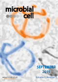
Cover: Fluorescence microscopy of mitochondrial morphology in
bck1∆
cnc1∆ cells expressing constitutively activated Rho1 (
RHO1 G19V). The mitochondria predominantly exhibit interconnected reticular phenotype. Image by Katrina F. Cooper (Rowan University School of Osteopathic Medicine, NJ, USA); modified by MIC. The cover is published under the Creative Commons Attribution (CC BY) license.
Enlarge issue cover
Peering into the ‘black box’ of pathogen recognition by cellular autophagy systems
Shu-chin Lai and Rodney J Devenish
Reviews |
page 322-328 | 10.15698/mic2015.09.225 | Full text | PDF |
Abstract
Autophagy is an intracellular process that plays an important role in protecting eukaryotic cells and maintaining intracellular homeostasis. Pathogens, including bacteria and viruses, that enter cells can signal induction of selective autophagy resulting in degradation of the pathogen in the autolysosome. Under such circumstances, the specific recognition and targeting of the invading pathogen becomes a crucial step for the subsequent initiation of selective autophagosome formation. However, the nature of the signal(s) on the pathogen surface and the identity of host molecule(s) that presumably bind the signal molecules remain relatively poorly characterized. In this review we summarise the available evidence regarding the specific recognition of invading pathogens by which they are targeted into host autophagy pathways.
The many facets of homologous recombination at telomeres
Clémence Claussin and Michael Chang
Reviews |
page 308-321 | 10.15698/mic2015.09.224 | Full text | PDF |
Abstract
The ends of linear chromosomes are capped by nucleoprotein structures called telomeres. A dysfunctional telomere may resemble a DNA double-strand break (DSB), which is a severe form of DNA damage. The presence of one DSB is sufficient to drive cell cycle arrest and cell death. Therefore cells have evolved mechanisms to repair DSBs such as homologous recombination (HR). HR-mediated repair of telomeres can lead to genome instability, a hallmark of cancer cells, which is why such repair is normally inhibited. However, some HR-mediated processes are required for proper telomere function. The need for some recombination activities at telomeres but not others necessitates careful and complex regulation, defects in which can lead to catastrophic consequences. Furthermore, some cell types can maintain telomeres via telomerase-independent, recombination-mediated mechanisms. In humans, these mechanisms are called alternative lengthening of telomeres (ALT) and are used in a subset of human cancer cells. In this review, we summarize the different recombination activities occurring at telomeres and discuss how they are regulated. Much of the current knowledge is derived from work using yeast models, which is the focus of this review, but relevant studies in mammals are also included.
A single mutation in the 15S rRNA gene confers non sense suppressor activity and interacts with mRF1 the release factor in yeast mitochondria
Ali Gargouri, Catherine Macadré and Jaga Lazowska
Research Articles |
page 343-352 | 10.15698/mic2015.09.223 | Full text | PDF |
Abstract
We have determined the nucleotide sequence of the mim3-1 mitochondrial ribosomal suppressor, acting on ochre mitochondrial mutations and one frameshift mutation in Saccharomyces cerevisiae. The 15s rRNA suppressor gene contains a G633 to C transversion. Yeast mitochondrial G633 corresponds to G517 of the E.coli 15S rRNA, which is occupied by an invariant G in all known small rRNA sequences. Interestingly, this mutation has occurred at the same position as the known MSU1 mitochondrial suppressor which changes G633 to A. The suppressor mutation lies in a highly conserved region of the rRNA, known in E.coli as the 530-loop, interacting with the S4, S5 and S12 ribosomal proteins. We also show an interesting interaction between the mitochondrial mim3-1 and the nuclear nam3-1 suppressors, both of which have the same action spectrum on mitochondrial mutations: nam3-1 abolishes the suppressor effect when present with mim3-1 in the same haploid cell. We discuss these results in the light of the nature of Nam3, identified by [1] as the yeast mitochondrial translation release factor. A hypothetical mechanism of suppression by “ribosome shifting” is also discussed in view of the nature of mutations suppressed and not suppressed.
The MAPKKKs Ste11 and Bck1 jointly transduce the high oxidative stress signal through the cell wall integrity MAP kinase pathway
Chunyan Jin, Stephen K. Kim, Stephen D. Willis and Katrina F. Cooper
Research Articles |
page 329-342 | 10.15698/mic2015.09.226 | Full text | PDF |
Abstract
Oxidative stress stimulates the Rho1 GTPase, which in turn induces the cell wall integrity (CWI) MAP kinase cascade. CWI activation promotes stress-responsive gene expression through activation of transcription factors (Rlm1, SBF) and nuclear release and subsequent destruction of the repressor cyclin C. This study reports that, in response to high hydrogen peroxide exposure, or in the presence of constitutively active Rho1, cyclin C still translocates to the cytoplasm and is degraded in cells lacking Bck1, the MAPKKK of the CWI pathway. However, in mutants defective for both Bck1 and Ste11, the MAPKKK from the high osmolarity, pseudohyphal and mating MAPK pathways, cyclin C nuclear to cytoplasmic relocalization and destruction is prevented. Further analysis revealed that cyclin C goes from a diffuse nuclear signal to a terminal nucleolar localization in this double mutant. Live cell imaging confirmed that cyclin C transiently passes through the nucleolus prior to cytoplasmic entry in wild-type cells. Taken together with previous studies, these results indicate that under low levels of oxidative stress, Bck1 activation is sufficient to induce cyclin C translocation and degradation. However, higher stress conditions also stimulate Ste11, which reinforces the stress signal to cyclin C and other transcription factors. This model would provide a mechanism by which different stress levels can be sensed and interpreted by the cell.
Intracellular phase for an extracellular bacterial pathogen: MgtC shows the way
Audrey Bernut, Claudine Belon, Chantal Soscia, Sophie Bleves, Anne-Béatrice Blanc-Potard
Microreviews |
page 353-355 | 10.15698/mic2015.09.227 | Full text | PDF |
Abstract
Pseudomonas aeruginosa is an extracellular pathogen known to impair host phagocytic functions. However, our recent results identify MgtC as a novel actor in P. aeruginosa virulence, which plays a role in an intramacrophage phase of this pathogen. In agreement with its intracellular function, P. aeruginosa mgtC gene expression is strongly induced when the bacteria reside within macrophages. MgtC was previously known as a horizontally-acquired virulence factor important for multiplication inside macrophages in several intracellular bacterial pathogens. MgtC thus provides a singular example of a virulence determinant that subverts macrophages both in intracellular and extracellular pathogens. Moreover, we demonstrate that P. aeruginosa MgtC is required for optimal growth in Mg2+ deprived medium, a property shared by MgtC factors from intracellular pathogens and, under Mg2+ limitation, P. aeruginosa MgtC prevents biofilm formation. We propose that MgtC has a similar function in intracellular and extracellular pathogens, which contributes to macrophage resistance and fine-tune adaptation to the host in relation to the different bacterial lifestyles. MgtC thus appears as an attractive target for antivirulence strategies and our work provides a natural peptide as MgtC antagonist, which paves the way for the development of MgtC inhibitors.


