
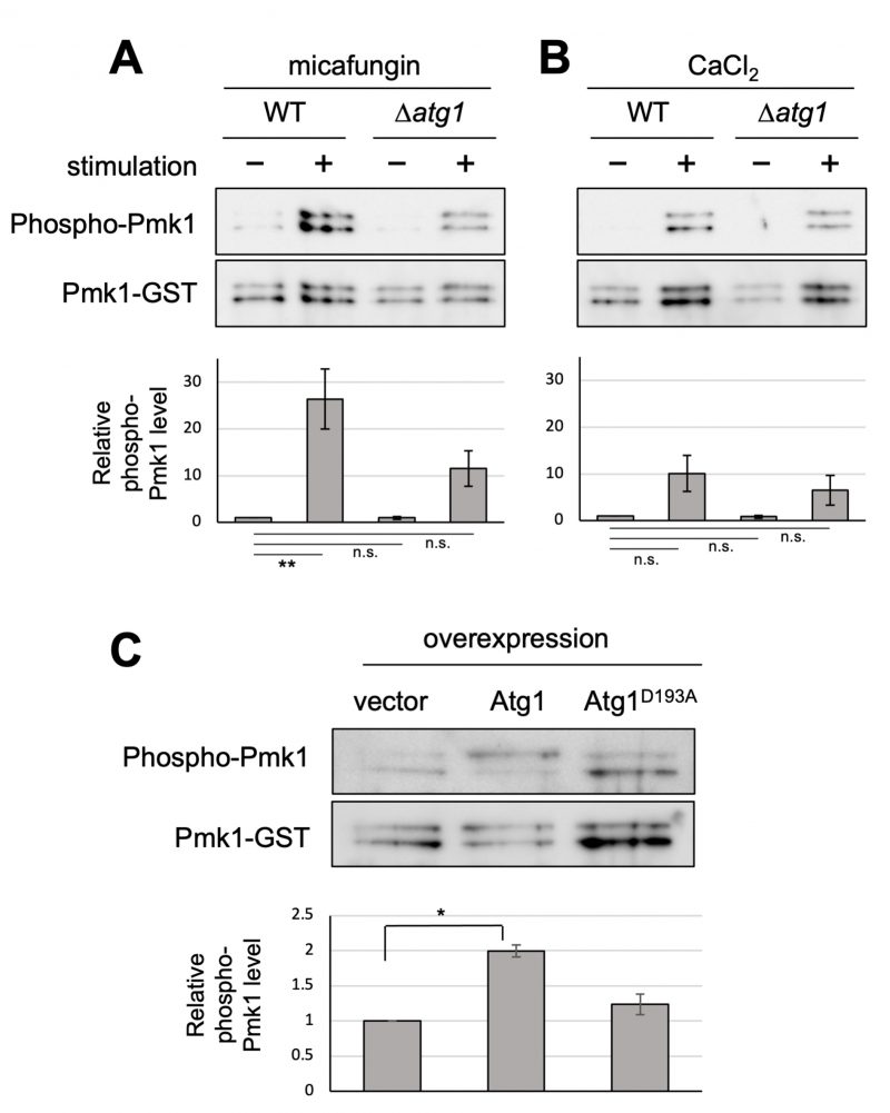
FIGURE 1: Atg1 facilitates the phosphorylation of Pmk1. (A and B) Impact of the deletion of atg1+ on Pmk1 phosphorylation. Wild-type (WT) and Δatg1 cells expressing the C-terminal GST-tagged Pmk1 from the endogenous pmk1 promoter were grown in EMM supplemented with 2 µg/ml micafungin (A) or with 200 mM CaCl2 (B) for 60 min at 27°C. Cell lysates bound to glutathione beads were immunoblotted with anti-phospho-ERK antibodies and anti-GST antibodies to detect phosphorylated Pmk1 and Pmk1-GST (loading control), respectively. Upper panel: representative immunoblot. Lower panel: Relative quantification of phosphorylated Pmk1 normalized by Pmk1-GST. Bar graphs show relative values to WT cells grown in the absence of micafungin nor CaCl2 as the mean ± standard error of the mean (SEM) of five independent experiments. N = 5; **p < 0.01 as assessed by a one-way ANOVA followed by the Dunnett's test for multiple comparisons. n.s. not significant. (C) Impact of overexpression of atg1+ on the Pmk1 phosphorylation. Δatg1 cells expressing Pmk1-GST under the endogenous pmk1 promoter and harboring pREP1-GFP, pREP1-atg1+-GFP, or pREP1-atg1D193A-GFP were grown in EMM without thiamine for 20 hr at 27°C. Cell lysates bound to glutathione beads were immunoblotted with anti-phospho-Pmk1 and anti-GST antibodies and quantified as described in (B). Bar graphs represent the mean ± SEM (N = 3; *p < 0.05 as assessed by Welch's two sample t-test). The levels of overexpression of WT Atg1 and kinase dead Atg1 were approximately equal (Figure S1).