
Back to article: Up-regulation of Osh6 boosts an anti-aging membrane trafficking pathway toward vacuoles
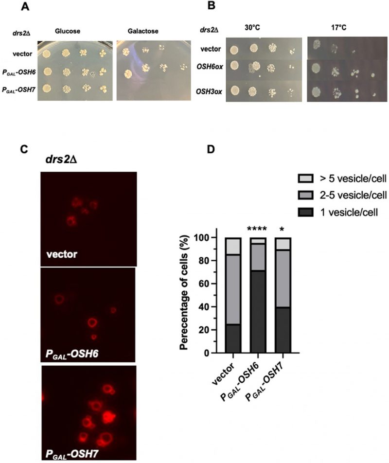
FIGURE 1: OSH6 genetically interacts with DRS2. (A) Growth of drs2Δ cells with vector, PGAL–OSH6 (pCB248), or PGAL–OSH7 (pCB247) on SC-URA with glucose or galactose media at 30°C for two days. (B) Growth of drs2Δ cells with vector, high copy OSH6 (pCB237), or high copy OSH3 (pCB238) on SC-URA at 30°C for two days or 17°C for ten days. For A and B, 5 µl of serially diluted cells (0.1 OD/ml for the left) were spotted on the plate and then incubated. (C) Vacuolar morphology of drs2Δ cells with vector or the indicated plasmid. Overnight cultures were labeled with FM4-64 for one hour and chased at 30°C for three hours and then photographed. (D) Quantitative analyses of vacuolar morphology from Fig. 1C. Cells were divided into three categories based on the number of vacuolar vesicles/cell. Sample sizes are 138 for drs2Δ (vector), 180 for drs2Δ (Pgal-PGAL–OSH6) and 194 for drs2Δ (PGAL–OSH7). A one-way ANOVA analysis shows that the fraction of cells with one vesicle/cell of PGAL–OSH6 is significantly different from that of vector (p<0.0001). Differences between PGAL–OSH7 and wild type is also significant (p=0.012).